![]()
86-551-67709456 (Working time)
86-13965027700 (Nonworking time)
Categories
Latest blog
Intraoperative radiotherapy is a kind of comprehensive therapy which is combined with local radiotherapy in the process of surgical treatment of malignant tumors. Its advantage is to treat the tumor layer after radical resection, the tumor node after reduction operation and the involved tissue nearby or the malignant tumor which can not be removed by operation. A special light limiting tube is used to aim at the lesion site for a single large dose irradiation, which not only prevents the gastrointestinal reaction of conventional irradiation, but also can kill or control the growth of tumor tissue.
Radiobiology
Abe's study in Japan shows that the retroperitoneal structure is more tolerant to single high dose irradiation. The National Cancer Institute (NCI) observed the effect of intraoperative radiotherapy with electron beam on normal tissues such as aorta, vena cava, kidney, ureter, bile duct and retroperitoneal soft tissue, blood vessels and intestinal anastomoses. The results showed that the blood vessel had a strong tolerance to general intraoperative radiotherapy, and the tissue structure of blood vessel had no significant change within 2 years after radiotherapy. Retroperitoneal structure can show fibrosis and scar to some extent. These reactions are small for high dose radiation. In contrast, the tolerance of functional organ system to radiation is poor. The table shows the tolerance of normal tissues and organs to intraoperative radiotherapy. It is important to protect the functional organ system during intraoperative radiotherapy.
Attached table: tolerance of normal tissue to intraoperative radiotherapy (NCI) in dogs
|
Tissue |
Maximum tolerable dose (Gy) |
Tissue effect |
|
Great vessels |
50 |
>30Gy Tubal fibrosis |
|
Kidney |
<20 |
Atrophy and fibrosis |
|
Ureter |
20 |
Fibrosis and stenosis |
|
Bile duct |
20 |
Fibrosis and stenosis |
|
Small intestine |
20 |
≥20Gy Ulcer, fibrosis, stenosis ≥30Gy Infarction, perforation |
|
Colon |
45 |
≥45Gy Ulcer, fibrosis, stenosis ≥50Gy perforation |
Treatment techniques and methods:
The disadvantages of atmospheric X-ray are poor penetration, insufficient deep dose and high shallow dose. Moreover, the absorption of bone is high, which may cause osteoporosis or malignant change. The advantage of atmospheric pressure X-ray machine is that it is cheap and can be used as a special machine for intraoperative radiotherapy, and can be placed in the operating room. The special designed light limiting tube must be used for intraoperative radiotherapy, which can be directly inserted into the surgical incision to contact the tumor area, so as to prevent radiation leakage and normal tissue sliding in. The light limiting cylinder is made of transparent plexiglass, which is convenient to observe the illuminated tissue directly. According to the location and size of the tumor, it can be made into a variety of different shapes, such as circle, rectangle, ellipse, polygon, etc., and can also be made into inclined.
In recent years, radiotherapy has been paid more attention in surgery. There are 150 research units of intraoperative radiotherapy in the world, with thousands of cases. The concept of treatment has developed from simple surgical resection to comprehensive treatment of surgery, radiotherapy, chemotherapy and immunotherapy.

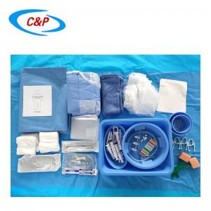
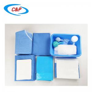
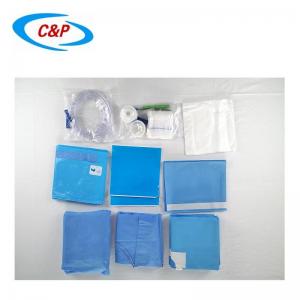
C&P medical radiology drape kits are made of absorbent and impermeable materials that help keep the work area dry and protect the patient. The customized nonwoven radiology packs are used to limit contamination of the surgical wound, the surgical instruments and the surgeon's hands.
Get in touch


Scan to wechat: